The applicability of LEXSY for structural biology was demonstrated by successful 15N-HSQC NMR analysis of a 28 kDa 15N-Val labeled protein purified from recombinant LEXSY strain grown in a synthetic LEXSY cultivation medium. All 18 Val residues of in vivo labeled protein could be completely assigned in 15N-HSQC NMR spectrum in full agreement with X-ray crystallography (Niculae et al. 2006). Since Leishmania tarentolae is auxotrophic for 11 amino acids and can be grown in chemically defined media multiple options for labeling strategies are offered. Alternatively to chemically defined media labeling strategies in complex media were developed (Foldynová-Trantírková et al. 2009).
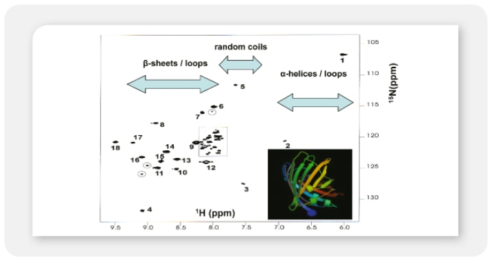
15N-HSQC NMR analysis of 15N-Val labeled EGFP purified from recombinant LEXSY strain. For detailed description refer to Niculae et al. (2006).
It was shown, that LEXSY-expressed proteins can be subjected successfully to crystallography and X-ray analysis. The resolution of a new protein structure was achieved for LEXSY expressed hu Cu/Zn superoxide dismutase SOD1 (Gazdak et al. 2010).
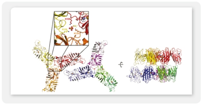
Structure determination of the new P212121 crystal form of LEXSY-produced human Cu/Zn superoxide dismutase (SOD1). The asymmetric unit contains six SOD dimers arranged as two triangular wheels around sulfate ions. The wheels are arranged in a side-to-side fashion (Gazdag et al. 2010).
Another example of a LEXSY produced protein which was successfully crystallized is the cysteine proteinase legumain (Dall & Brandstetter 2012). Screening for crystallization conditions of legumain has been carried out with the JBScreen Classic kit. Super-activated legumain was successfully crystallized in complex with a specific inhibitor and using the heavy-atom soaking method, and mercury-derivative crystals were obtained. Single anomalous scattering dataset was collected to a resolution of 2.47 Å and the structure determined with a combination of isomorphous and anomalous scattering. The preliminary X-ray diffraction data analysis of active legumain has been presented and the structure of legumain glycoforms will be published elsewere (Hans Brandstetter, personal communication).
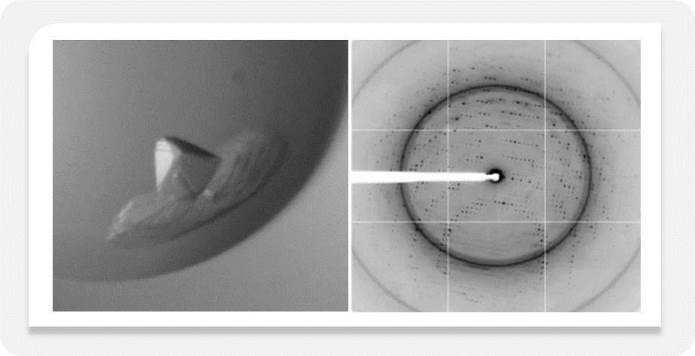
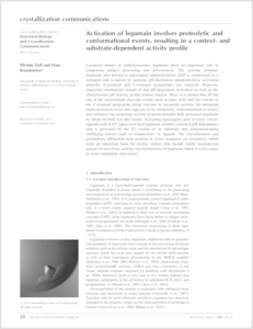
Crystals of mature legumain produced in LEXSY and X-ray diffraction image (from Dall & Brandstetter 2012).