Existing tools for the visualization of mitochondrial DNA (mtDNA) have significant limitations. Consequently, the formation and regulation of mtDNA in cells is not fully understood[1].
Currently, mtDNA is visualized with EdU (5-ethynyl-dU). However, EdU-based mtDNA-labeling causes significant nuclear DNA staining which can only be partially overcome by aphidicolin treatment[1].
Selective staining can now be achieved using PdG (6-O-Propynyl-dG). Visualization of such azide-functionalized fluorescent dyes is achieved via the Copper(I)-catalyzed azide-alkyne cycloaddition (CuAAC) (Fig. 1) with the added benefit of lower cytotoxicity compared to EdU[2,3].
Cellular incorporation of PdG into replicating mtDNA resulted in a clear, almost selective mitochondrial staining, as shown by co-localization with cytochrome c. No nuclear staining is seen in contrast with EdU (PdG does not cause fluorescence in the DAPI stained region) (Fig. 1B)[2,3].
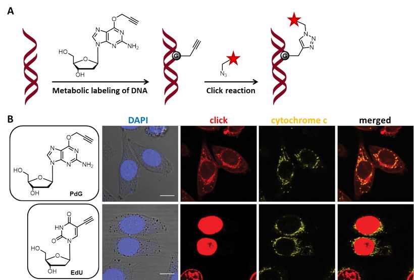
Figure 1: A) Incorporated PdG in mtDNA is visualized with an azide-functionalized fluorescent dye via CuAAC. B) Comparison of specificity in mtDNA labeling with PdG and EdU. HeLaP4 cells were treated with PdG or EdU for 7 h. The cells were then fixed and click stained (red) together with a nuclear (DAPI, blue) and cytochrome c (yellow) staining. Confocal microscopy was used to acquire Z stacks covering the whole-cell volume. (Scale bar: 10 µm) (modified according to [2]).
[1] Jones et al. (2020) Visualizing, quantifying, and manipulating mitochondrial DNA in vivo. J. Biol. Chem. 295:17588.
[2] De Wit et al. (2019) Design of reverse transcriptase–specific nucleosides to visualize early steps of HIV-1 replication by click labeling. J. Biol. Chem. 294 (31):11863.
[3] Venkatesham et al. (2019) Propargylated Purine Deoxynucleosides: New Tools for Fluorescence Imaging Strategies. Molecules 24 (3):468.
