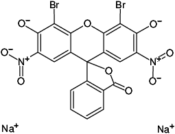
Eosin Scarlet
| Cat. No. | Amount | Price (EUR) | Buy / Note |
|---|---|---|---|
| CO-304 | 300 μl | 13,70 | Add to Basket/Quote Add to Notepad |
For general laboratory use.
Shipping: shipped at ambient temperature
Storage Conditions: store at ambient temperature
Shelf Life: 12 months
Molecular Formula: C20H6Br2N2Na2O9
Molecular Weight: 624.08 g/mol
CAS#: 548-24-3
EC number: 208-943-1
Applications:
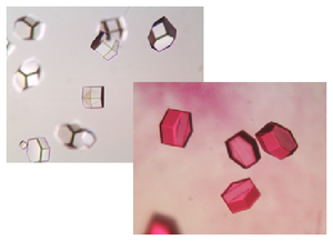
JBS Bright Red is a crystal dye used to stain macromolecular crystals, i.e. protein, peptide and nucleic acid crystals in order to differentiate them from small molecules and salt crystals.
Description:
Crystallization screening with high concentrations of precipitant and salt may lead to the formation of salt crystals. It is quite difficult to make a distinction between these false positives and true protein crystals.
Staining of crystals with appropriate dyes is a very straightforward method to differentiate between macromolecular crystals and salt crystals [1].
Protein and salt crystals differ substantially in their solvent content. Small crystal dyes, like JBS Bright Red, are able to permeate the solvent channels of a protein, thus coloring the protein red. In contrast, salt crystals are tightly packed and do not possess large solvent channels. They will therefore remain colourless.
Usage:
Simply add 0.5-1 μl of JBS Bright Red to the crystallization drop containing the crystals of interest.
Coloring Time:
JBS Bright Red colors protein crystals after a few minutes. Even if the color of the solution is only faintly red under the microscope, proteins will be stained within 5-15 min.
Very occasionally, it has been reported that protein crystals did not absorb crystal dyes [2].
BIOZ Product Citations:
Selected References:
[1] Wilkosz et al. (1995) Preliminary characterization of EcoRI-DNA co-crystals: incomplete factorial design of oligonucleotide sequences. Acta Cryst. D 51:938.
[2] Eckert et al. (2003) Crystallization and preliminary X-ray analysis of Alicyclobacillus acidocaldarius endoglucanase CelA. Acta Cryst. D 59:139.
 Powered by Bioz